
Contents
Retinal Detachment Treatment in Turkey
Retinal detachment is one of the most urgent conditions in ophthalmology. When the retina separates from the underlying supportive tissue, the affected photoreceptor cells lose their blood supply, and permanent vision loss can follow within hours or days. Turkey has established itself as a leading destination for retinal detachment treatment, combining internationally accredited eye hospitals, highly experienced vitreoretinal surgeons, and treatment costs that are a fraction of what patients pay in the US or UK. This guide covers everything you need to know about the procedure, the types of surgery available, expected costs, recovery, and how A-Medical can help you access the best care in Turkey quickly and affordably.
What Is Retinal Detachment?

The retina is a thin, light-sensitive layer of nerve tissue that lines the back wall of the eye. It captures incoming light and converts it into electrical signals that travel through the optic nerve to the brain, producing the images we see. The retina sits on top of the retinal pigment epithelium (RPE) and the choroid, a vascular layer that supplies it with oxygen and essential nutrients.
Retinal detachment occurs when the retina peels away from the RPE and choroid. Once separated, the retinal cells are cut off from their blood supply. Without prompt intervention, these cells begin to die, and the resulting vision loss can become irreversible. That is why retinal detachment is classified as an ophthalmic emergency that demands immediate evaluation and, in most cases, urgent surgical repair.
There are three clinically recognized types of retinal detachment:
- Rhegmatogenous retinal detachment: The most common form. A tear or hole in the retina allows vitreous fluid to seep underneath, gradually lifting the retina from the RPE. Age-related vitreous degeneration and posterior vitreous detachment (PVD) are the primary triggers.
- Tractional retinal detachment: Scar tissue or fibrovascular membranes on the retinal surface contract and physically pull the retina away from its bed. This type is most frequently associated with proliferative diabetic retinopathy and sickle cell disease.
- Exudative (serous) retinal detachment: Fluid accumulates beneath the retina without any tear or traction. Underlying causes include ocular tumors, severe uveitis, central serous chorioretinopathy, and certain vascular abnormalities.
Symptoms of Retinal Detachment

Retinal detachment itself is painless, which makes recognizing its warning signs critically important. Many patients describe a characteristic sequence of visual disturbances:
- A sudden increase in floaters, appearing as dark spots, threads, or cobweb-like shapes drifting across the visual field
- Bright flashes of light (photopsia), especially noticeable in dim lighting or at the periphery of vision
- A shadow or dark curtain spreading across part of the visual field
- Blurred or distorted central or peripheral vision
- A sudden, unexplained decrease in visual acuity
If you experience any combination of these symptoms, you should seek emergency ophthalmic evaluation immediately. Timely diagnosis is the single most important factor in preserving vision after a retinal detachment.
Causes and Risk Factors of Retinal Detachment

Understanding who is at risk helps both patients and referring physicians identify the condition early. The following factors significantly increase the likelihood of developing a detached retina:
- Age over 50: Posterior vitreous detachment (PVD) becomes increasingly common with age as the vitreous gel liquefies and shrinks, creating traction on the retina.
- High myopia (nearsightedness): An elongated eyeball stretches and thins the retina, predisposing it to tears and detachment.
- Previous eye surgery: Patients who have undergone cataract surgery or other intraocular procedures carry an elevated risk of retinal tears.
- Ocular trauma: A direct blow to the eye or face can create retinal tears.
- Diabetic retinopathy: Chronically elevated blood sugar damages retinal blood vessels and promotes scar tissue formation, leading to tractional detachment.
- Family history: A genetic predisposition to retinal weakness, lattice degeneration, or retinoschisis raises risk.
- Previous retinal detachment in the other eye: Having experienced a detachment in one eye significantly increases the probability of it occurring in the fellow eye.
- Peripheral retinal degeneration: Conditions such as lattice degeneration thin the peripheral retina and create predisposing lesions.
Who Is a Good Candidate for Retinal Detachment Treatment?

Almost every patient diagnosed with a retinal detachment is a candidate for treatment, because the alternative is progressive, irreversible blindness. However, the specific surgical approach and expected outcome depend on the individual case. Ideal candidates include:
- Patients with acute rhegmatogenous detachment, especially when the macula is still attached (macula-on), where rapid surgery offers the best chance of preserving central vision
- Individuals with retinal tears or holes that have not yet progressed to full detachment, who can benefit from preventive laser or cryotherapy
- Diabetic patients with tractional detachment threatening or involving the macula
- Patients whose previous retinal surgery has failed and require revision vitrectomy or combined procedures
- Patients in overall good general health who can tolerate anesthesia and comply with post-operative positioning requirements
Patients with significant systemic comorbidities, advanced proliferative vitreoretinopathy (PVR), or extremely long-standing detachments may still undergo surgery, but expectations regarding visual recovery should be realistic. Your vitreoretinal surgeon in Turkey will assess each case individually and recommend the most appropriate intervention.
How Retinal Detachment Is Diagnosed
Accurate diagnosis requires a thorough ophthalmic examination. In Turkey's leading eye hospitals, the diagnostic workup typically includes:
- Dilated fundus examination: Using indirect ophthalmoscopy with scleral depression, the retina specialist inspects the entire retina for tears, holes, and areas of detachment.
- Optical coherence tomography (OCT): High-resolution cross-sectional imaging of the retinal layers provides detailed visualization of the detachment and macular status.
- B-scan ultrasonography: When vitreous hemorrhage or dense cataracts prevent direct visualization, ultrasound confirms the presence and extent of detachment.
- Fundus fluorescein angiography (FFA): In selected cases, FFA evaluates retinal blood flow and identifies vascular abnormalities contributing to the detachment.
- Visual field testing and visual acuity assessment: Baseline measurements help guide treatment urgency and set realistic expectations for post-surgical recovery.
Types of Retinal Detachment Treatment in Turkey

Turkey's accredited eye clinics offer the full spectrum of retinal detachment repair techniques. The choice of procedure depends on the type, severity, and location of the detachment, as well as the presence of complicating factors such as PVR or vitreous hemorrhage.
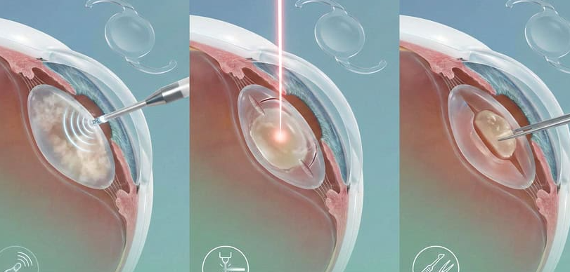
Laser Surgery (Photocoagulation)
Laser photocoagulation is a non-invasive, office-based procedure used to treat retinal tears and small, localized areas of shallow detachment before the condition progresses. The ophthalmologist directs a focused laser beam at the tissue surrounding the tear, creating controlled thermal burns that form scar tissue. This scar acts like a weld, sealing the retina to the RPE and preventing fluid from migrating underneath. The procedure takes 15 to 30 minutes, requires only topical anesthetic eye drops, and patients can usually return to normal activities within a day. Laser treatment is most effective as a preventive measure; once a large area of the retina has fully detached, laser alone is insufficient. For patients exploring laser eye procedures in Turkey, this approach represents one of the most accessible entry points into retinal care.
Cryotherapy (Cryopexy)
Cryotherapy works on a similar principle to laser photocoagulation but uses extreme cold instead of heat. A freezing probe (cryoprobe) is applied to the outer surface of the eye directly over the retinal tear. The resulting freeze-thaw cycle causes localized inflammation and scarring that seals the tear. Cryotherapy is particularly useful when the tear is located far in the periphery of the retina, where laser access may be limited, or when media opacities such as vitreous hemorrhage obscure the laser's path. Like laser treatment, cryopexy is most effective for tears that have not yet caused significant detachment.
Pneumatic Retinopexy
Pneumatic retinopexy is a minimally invasive procedure that can often be performed in an outpatient clinic under local anesthesia. The surgeon injects a small gas bubble (usually sulfur hexafluoride or perfluoropropane) into the vitreous cavity. The patient is then positioned so that the bubble floats up against the retinal tear, pushing the detached retina back into contact with the underlying tissue. Once the retina is repositioned, laser photocoagulation or cryopexy is applied around the tear to create a permanent seal. The gas bubble gradually absorbs on its own over one to eight weeks. Pneumatic retinopexy is best suited for uncomplicated, single-break rhegmatogenous detachments in the upper portion of the retina. Patients must maintain a specific head position for several days, which is a key factor in treatment success.
Vitrectomy Surgery
Vitrectomy is the most commonly performed surgery for complex retinal detachments today. Using modern 25-gauge or 27-gauge microincision techniques, the surgeon makes tiny incisions in the sclera and removes the vitreous gel that is pulling on the retina. Subretinal fluid is drained, and laser or cryotherapy is applied around every identified tear. At the end of the procedure, the vitreous cavity is filled with a gas bubble or silicone oil tamponade to hold the retina in position while it heals. The gas bubble reabsorbs naturally over weeks; silicone oil may need to be removed in a secondary procedure months later. Vitrectomy is indicated for macula-off detachments, cases with multiple or giant tears, tractional detachments from diabetic retinopathy, detachments complicated by PVR, and recurrent detachments after prior surgery. In experienced hands, pars plana vitrectomy achieves anatomical reattachment rates of 85% to 95% with a single operation.
Scleral Buckling Surgery
Scleral buckling is a time-tested external approach to retinal detachment repair. The surgeon sutures a piece of silicone sponge or solid silicone band (the buckle) onto the outer wall of the eye at the location of the retinal break. This indents the sclera inward, reducing vitreous traction and bringing the eye wall into contact with the detached retina. Cryopexy is applied to the area of the break to promote adhesion. In many cases, subretinal fluid is drained through a small external incision to facilitate retinal reattachment. The buckle remains permanently in place and is not visible externally. Scleral buckling is highly effective for uncomplicated rhegmatogenous detachments, particularly in younger, phakic patients, and can be combined with vitrectomy for more complex cases.
How Retinal Detachment Surgery Is Performed
The surgical workflow for retinal detachment treatment in Turkey follows internationally standardized protocols. Before surgery, the patient undergoes a comprehensive ophthalmic evaluation, including OCT, B-scan ultrasound, and blood work. The procedure is typically performed under local anesthesia (retrobulbar or peribulbar block), though general anesthesia may be used for pediatric patients or lengthy combined procedures.
During vitrectomy, for example, the surgeon inserts three micro-cannulas through the sclera: one for an infusion line that maintains intraocular pressure, one for a fiberoptic light source, and one for the vitreous cutter and other instruments. Under high-magnification visualization using a wide-angle viewing system, the surgeon carefully removes vitreous gel, peels membranes, identifies and treats all retinal breaks, drains subretinal fluid, and instills an appropriate tamponade agent. The entire procedure typically lasts 45 minutes to two hours depending on complexity.
Post-operatively, the patient may be required to maintain face-down positioning for several days to allow the gas or oil tamponade to press effectively against the reattached retina. Anti-inflammatory and antibiotic eye drops are prescribed, and follow-up appointments are scheduled at regular intervals to monitor healing.
Recovery After Retinal Detachment Treatment
Recovery timelines vary depending on the procedure performed, the extent of the detachment, and individual healing responses. After vitrectomy with gas tamponade, patients may need to maintain a specific head position for five to ten days. Vision will be blurry while the gas bubble is present but gradually improves as the bubble shrinks and is replaced by the eye's natural aqueous humor over two to eight weeks.
For scleral buckling, the eye may be swollen and sore for several weeks, but most patients can return to light daily activities within one to two weeks. Flying or traveling to high altitudes is prohibited while a gas bubble is in the eye, as changes in atmospheric pressure can cause the bubble to expand dangerously.
Full visual recovery can take three to six months, and in some cases up to a year. Patients should expect regular follow-up visits during this period. Vision improvement depends heavily on whether the macula was attached (macula-on) or detached (macula-off) at the time of surgery. Macula-on cases generally achieve better visual outcomes.
Risks and Success Rates of Treatment
Modern retinal detachment surgery is highly successful. A single vitrectomy or scleral buckle procedure achieves anatomical reattachment in approximately 85% to 95% of cases. When re-operation is included, final reattachment rates exceed 95%. However, anatomical success does not always equal full visual recovery, particularly when the macula was detached prior to surgery.
Potential risks and complications include:
- Cataract formation (especially after vitrectomy in phakic patients)
- Elevated intraocular pressure (glaucoma)
- Re-detachment, which occurs in approximately 5% to 10% of cases, most often within the first three months
- Proliferative vitreoretinopathy (PVR), where scar tissue forms on the retinal surface and causes recurrent traction
- Endophthalmitis (intraocular infection), which is rare but serious
- Vitreous hemorrhage during or after surgery
Turkey's leading retina centers mitigate these risks through advanced surgical instruments (25- and 27-gauge micro-incision systems), high-definition 3D visualization platforms, and rigorous post-operative monitoring protocols including regular OCT imaging and fundus examinations.
Cost of Retinal Detachment Treatment in Turkey (2026)
One of the most compelling reasons international patients choose Turkey for retinal detachment treatment is the significant cost advantage. Below is an overview of typical prices for different procedures in Turkey:
- Laser photocoagulation: $500 to $1,000
- Pneumatic retinopexy: $1,500 to $2,750
- Scleral buckling: $1,800 to $2,800
- Vitrectomy (pars plana): $2,500 to $4,000
- Combined procedures (vitrectomy + scleral buckle or silicone oil): $4,000 to $6,000
These prices at Turkish hospitals typically include pre-operative diagnostics, surgeon and anesthesia fees, surgical materials, hospital stay, and initial post-operative consultations. Many clinics also bundle accommodation, airport transfers, and interpreter services into their medical tourism packages.
Retinal Detachment Treatment Costs in the USA
In the United States, the cost of retinal detachment repair is substantially higher. A standard pars plana vitrectomy typically ranges from $8,000 to $20,000 per eye, depending on facility type, geographic location, and case complexity. Pneumatic retinopexy, while less expensive, still costs between $5,000 and $10,000. Scleral buckling falls in the $7,500 to $15,000 range. Importantly, US pricing often covers only the surgeon's fee and facility charges; anesthesia, diagnostic imaging, and post-operative care may be billed separately, driving total costs even higher. For the full pricing picture on eye surgery costs across countries, consult our dedicated guide.
Retinal Detachment Treatment Costs in the UK
Private retinal detachment treatment in the United Kingdom typically costs between £5,000 and £8,000 (approximately $6,300 to $10,100) per eye. London-based clinics may charge more, with vitrectomy procedures starting from around £7,000. Although NHS treatment is available at no direct cost, waiting times for non-emergency referrals can be lengthy, which is a critical concern given the time-sensitive nature of retinal detachment.
Why Turkey Offers Better Value
When comparing Turkey with the USA and the UK, patients can save 60% to 85% on their total treatment costs without any compromise in surgical quality. Turkish ophthalmology hospitals hold JCI accreditation, ISO certifications, and Ministry of Health accreditation. Surgeons use the same micro-incision vitrectomy systems, wide-angle viewing platforms, and tamponade agents available in the world's top-tier centers. The cost savings are primarily driven by lower operational overheads, favorable exchange rates, and government-supported medical tourism incentives, not by any reduction in clinical standards or technology.
Why Choose Turkey for Retinal Detachment Treatment?
Advanced Eye Clinics and Technology
Turkey is home to a dense network of state-of-the-art eye hospitals in Istanbul, Ankara, Izmir, and Antalya. These facilities are equipped with the latest generation of high-speed vitrectomy machines, intraoperative OCT systems, 3D visualization microscopes, and multi-wavelength laser platforms. Many have received international accreditation from bodies such as JCI, TEMOS, and TUV NORD.
Experienced Retina Specialists
Turkish vitreoretinal surgeons are recognized worldwide for their expertise. Many have trained or completed fellowships at prestigious institutions such as Moorfields Eye Hospital in London, the Doheny Eye Institute in Los Angeles, and leading European university clinics. Some of the most distinguished professors in the field have personally performed over 10,000 retinal procedures. This depth of experience is directly reflected in Turkey's high surgical success rates.
High Success Rates in Emergency Eye Care
Turkish eye clinics report retinal reattachment success rates of approximately 90% with a single operation, consistent with the best centers globally. For cases requiring reoperation, final success rates exceed 95%. The emphasis on rapid diagnosis, immediate surgical availability, and comprehensive post-operative follow-up contributes significantly to these outcomes.
Affordable Treatment Options
Retinal detachment treatment in Turkey costs 60% to 85% less than equivalent procedures in the United States and 50% to 70% less than in the UK or Western Europe. All-inclusive packages offered by Turkish clinics frequently cover the surgery, hospital stay, diagnostics, medications, hotel accommodation, airport transfers, and personal translation services, providing exceptional value for international patients.
Medical Tourism Support Services
Turkey treated over 1.2 million international patients in 2025, and the country's medical tourism infrastructure is among the most developed in the world. From dedicated international patient departments at major hospitals to multilingual coordinators and digital telemedicine consultations, the entire journey is designed for seamless cross-border healthcare delivery.
Best Clinics for Retinal Detachment Treatment in Turkey
Turkey is home to numerous world-class eye hospitals with dedicated retina departments. Below are some of the top facilities where international patients receive retinal detachment surgery:
Dunyagoz Hospital (World Eye Hospital)
Dunyagoz is Turkey's largest and most well-known eye hospital group, with multiple branches across Istanbul and other cities. The hospital's Retina and Vitreous Department is led by professors with over 12,000 retinal procedures to their name, including Prof. Dr. Hamdi Er, a member of the American Society of Retina Specialists (ASRS) and the European Vitreoretinal Society (EVRS). Dunyagoz holds TUV NORD ISO 9001 certification and treats over 40,000 international patients annually. The clinic offers comprehensive vitrectomy, scleral buckling, and laser photocoagulation services, with all-inclusive international patient packages that cover surgery, accommodation, transfers, and interpreter support.
Memorial Sisli Hospital
Memorial Sisli Hospital in Istanbul holds JCI accreditation and has been a pioneer in Turkish healthcare since its founding. The hospital's Ophthalmology Department features advanced vitreoretinal surgeons such as Prof. Dr. Abdullah Ozkaya, a member of the European Vitreoretinal Society with over two decades of experience. Memorial Sisli was Turkey's first LEED Platinum-certified healthcare facility and has treated patients from over 92 countries. Vitrectomy packages at Memorial typically include pre-operative diagnostics, surgery, post-operative consultations, and coordination services.
Anadolu Medical Center
Affiliated with Johns Hopkins Medicine, Anadolu Medical Center in Istanbul's Gebze district combines American healthcare standards with Turkish surgical expertise. The Ophthalmology Department's retina team uses intraoperative OCT-guided vitrectomy systems for maximum precision. Anadolu is JCI-accredited and emphasizes a multidisciplinary approach to complex retinal cases, collaborating with vitreoretinal specialists, diabetologists, and neurologists when necessary.
Hisar Intercontinental Hospital
Hisar Intercontinental Hospital in Istanbul holds JCI accreditation and houses an experienced vitreoretinal surgery team. Dr. Omer Kocabiyik, who has performed over 1,100 retinal procedures, leads the retina unit. The hospital offers all-inclusive retinal detachment surgery packages that typically cover surgery, anesthesia, OCT scans, hospital stay, follow-up consultations, and transportation. Hisar is particularly popular among international patients from Europe and the Middle East.
How A-Medical Can Help?
Planning emergency eye surgery abroad can feel overwhelming, especially when every day counts. A-Medical eliminates the complexity by serving as your trusted partner from the first inquiry to your final follow-up appointment. Here is what we provide:
- No waiting lists: We secure priority appointments with Turkey's top vitreoretinal surgeons, often within 24 to 48 hours of your initial contact.
- Best clinic and doctor matching: Based on your diagnosis, medical history, and treatment goals, we match you with the most suitable hospital and surgeon for your specific case.
- Accommodation arrangements: We organize comfortable hotel stays near your treatment center, with options ranging from budget-friendly to premium.
- Professional interpreter services: A dedicated medical interpreter accompanies you to every appointment, ensuring clear communication with your healthcare team.
- Airport and local transfers: All ground transportation, from airport pick-up to hospital visits and back, is arranged for your comfort and convenience.
- Most affordable pricing: Through our established partnerships with Turkey's leading eye hospitals, we help you access the most competitive treatment costs available.
If you or a loved one has been diagnosed with retinal detachment and you are considering treatment in Turkey, contact A-Medical today for a free consultation and personalized treatment plan.
Conclusion
Retinal detachment is a sight-threatening emergency, but with timely and expert surgical intervention, vision can be preserved and often significantly restored. Turkey offers an unmatched combination of advanced surgical technology, world-class vitreoretinal surgeons, internationally accredited hospitals, and treatment costs that are a fraction of what patients pay in the US or UK. Whether you require laser treatment for a retinal tear, pneumatic retinopexy for a localized detachment, or complex vitrectomy for a macula-off case, Turkish eye clinics deliver outcomes that rival the best centers anywhere in the world.
Retinal detachment treatment in Turkey through A-Medical gives you the added advantage of fast-tracked appointments, personalized clinic matching, accommodation, interpreter services, and ground transportation, so you can focus entirely on your recovery. Reach out to A-Medical today to receive a free, no-obligation treatment plan and take the first step toward restoring your vision.
Frequently Asked Questions
Is retinal detachment an emergency?
Yes, retinal detachment is an ophthalmic emergency. The retina loses its blood supply once detached, and the photoreceptor cells begin to die. Without prompt surgical intervention, vision loss becomes permanent.
How soon should retinal detachment treatment be done?
Treatment should be performed as soon as possible after diagnosis. In macula-on cases, surgery is ideally done within 24 hours to prevent the detachment from reaching the central vision area. Even in macula-off cases, earlier surgery is associated with better visual outcomes.
What is the success rate of retinal detachment surgery?
A single vitrectomy or scleral buckle achieves anatomical reattachment in 85% to 95% of cases. With reoperation if needed, final success rates exceed 95%. Visual recovery depends on factors such as macular involvement and the duration of detachment.
Will vision fully recover after retinal detachment treatment?
Visual recovery varies. Patients with macula-on detachments who receive timely surgery often recover excellent vision. When the macula has been detached, some degree of permanent visual impairment is common, though meaningful improvement is still achievable. Full visual stabilization may take up to 12 months.
Can retinal detachment recur after surgery?
Recurrence occurs in approximately 5% to 10% of cases, usually within the first three months. Risk factors include multiple retinal breaks, PVR, and extensive lattice degeneration. Close post-operative monitoring with regular OCT and fundus examinations allows early detection and prompt re-intervention.
Is retinal detachment surgery painful?
No, the surgery itself is painless because it is performed under local or general anesthesia. After the procedure, patients may experience mild discomfort, a foreign-body sensation, or slight swelling, which is managed effectively with prescribed eye drops and anti-inflammatory medications.


