Treatment Abroad – Find Verified Clinics, Reviews & Prices
Treatment Abroad made simple by A-Medical. Explore top clinics worldwide and start your journey toward better health today
We make healthcare abroad
simple, transparent, and fully guided from start to finish
Submit Your Request
Tell us about your treatment needs. We carefully review your case and match you with the most suitable hospitals and doctors.
Get Official Price Quotes
We provide verified treatment offers and proformas directly from hospitals — no middlemen, full transparency.
Connect with Doctors
We arrange direct calls or video consultations with hospitals and doctors so you can discuss your treatment plan confidently.
Confirm & Start Treatment
Once you accept the treatment plan, we schedule your hospital appointment and support you throughout your medical journey.
Clinics
Discover our network of world-class medical facilities offering comprehensive healthcare services
Why Choose Us
With us, you are in safe hands. We focus on quality, transparency, and patient-first care. Here’s what sets us apart
Only Accredited Hospitals
We partner exclusively with internationally accredited and top-ranked hospitals.
Direct & Transparent Payments
You pay directly at the hospital, with no hidden fees.
Free Direct Call & Video Consultation
Speak with our experts and doctors before making any decisions.
Best Doctor & Hospital Match
We carefully select the most suitable specialist and clinic for your treatment needs.
Worldwide Hospital Network
Access to a vast global network across 90+ countries.
More Affordable Pricing
We negotiate better prices from hospitals than you would get by contacting them directly.
Medical Conditions
Explore our comprehensive coverage of medical conditions and find the right treatment options

detoxification
The physiological or medical process of removing toxic substances from a living organism, primarily carried out by the liver.

Glioma
A glioma is a type of brain tumor that develops in the central nervous system and originates from glial cells. These cells play a vital role in maintaining the health and function of nerve cells. When genetic mutations occur in glial cells, uncontrolled growth can lead to tumor formation. Gliomas can develop in different parts of the brain and spinal cord. The most common locations include the cerebral hemispheres, brainstem, cerebellum, and spinal cord. The tumor’s size, location, and growth rate vary significantly from one patient to another. Although gliomas are relatively rare compared with other cancers, some subtypes are more common than others. Understanding glioma types, symptoms, diagnosis, and treatment options is essential for early detection and effective care. Specialized treatment is available at leading centers such as Sheba Medical Center. Glioma Types Glioma is an umbrella term that includes several subtypes, classified based on the type of glial cell involved and the tumor’s location. Astrocytoma Astrocytomas arise from astrocytes, star-shaped glial cells that support and protect neurons. These tumors often infiltrate healthy brain tissue, making high-grade astrocytomas difficult to remove completely. Grades: Grade 1: Pilocytic astrocytoma, SEGA, pleomorphic xanthoastrocytoma Grade 2: Diffuse astrocytoma Grade 3: Anaplastic astrocytoma Grade 4: Glioblastoma Most diagnosed gliomas are astrocytomas, ranging from low-grade to highly aggressive forms. Brainstem Glioma Brainstem gliomas develop in the brainstem, a region that controls vital functions such as breathing and heart rate. Because of their location, surgery is often not possible. Types: Focal brainstem gliomas: Slow-growing, localized, less aggressive Diffuse intrinsic pontine glioma (DIPG): Rapidly growing, highly aggressive, infiltrates surrounding tissue Ependymoma Ependymomas originate from ependymal cells, which line the ventricles of the brain and the central canal of the spinal cord. Grades: Grade 1–2: Low-grade, often found in the spinal cord Grade 3: High-grade, more common in the brain Mixed Glioma Mixed gliomas contain more than one type of glial cell. Their behavior and aggressiveness depend on the specific cell combination and tumor grade. Oligodendroglioma Oligodendrogliomas arise from oligodendrocytes, cells that insulate nerve fibers. Grades: Grade 2: Slow-growing, low-grade Grade 3 (Anaplastic): Fast-growing, aggressive Optic Pathway Glioma Optic gliomas develop along the optic nerve, optic chiasm, or hypothalamus, often affecting vision. Stages: Stage 1: Optic nerve involvement Stage 2: Optic chiasm involvement Stage 3: Hypothalamic involvement Glioma Symptoms Symptoms depend on tumor location and size. Common signs include: Persistent or worsening headaches Difficulty with balance and walking Vision problems Nausea and vomiting Sudden seizures Confusion or memory problems Personality or behavioral changes Speech difficulties Unexplained weight changes Symptoms vary widely, and not all patients experience the same signs. Risk Factors Known factors associated with a higher risk of glioma include: Genetic conditions: Neurofibromatosis type 1, Turcot syndrome, Li-Fraumeni syndrome Family history: Rare, but reported in some families Previous exposure to ionizing radiation, especially childhood radiotherapy Age: More common in children aged 5–8 and adults aged 45–65 Glioma Diagnosis Diagnosis involves multiple steps: Physical exam and medical history Neurological examination CT scan for structural imaging MRI for detailed tumor visualization Biopsy to identify tumor type and grade Lumbar puncture (in select cases) Molecular testing to guide personalized treatment Glioma Grading Gliomas are graded from 1 to 4, based on how abnormal the tumor cells appear and how fast they grow. Low-Grade Gliomas (Grades 1–2) Slow-growing Less aggressive Often treated with surgery ± chemotherapy Generally favorable prognosis High-Grade Gliomas (Grades 3–4) Rapid growth Highly malignant May arise spontaneously or from lower-grade tumors Require aggressive treatment Glioma Treatment Options Treatment depends on tumor type, grade, and location: Surgery Radiation therapy Chemotherapy High-dose chemotherapy with bone marrow transplant (select cases) Targeted therapy, which attacks cancer cells while sparing healthy tissue Glioma Prognosis (Approximate 5-Year Survival Rates) Grade 1: ~95% Grade 2: ~40–50% Grade 3: ~25–30% Grade 4: ~5–10% Prognosis varies based on individual factors and response to treatment.

Lacunar syndromes
Lacunar Syndromes – Clear Clinical Overview Lacunar syndromes are a group of distinct neurological presentations caused by small deep infarcts (lacunes) in the brain. These infarcts result from occlusion of small penetrating arteries, most commonly due to chronic hypertension, but also diabetes and smoking. Unlike large strokes, lacunar strokes do not involve the cerebral cortex, which explains their characteristic pattern of symptoms. The Five Classical Lacunar Syndromes 1. Pure Motor Stroke (Pure Motor Hemiparesis) Most common lacunar syndrome Weakness or paralysis of the face, arm, and leg on one side No sensory loss Common lesion sites: Posterior limb of the internal capsule Basis pontis 2. Pure Sensory Stroke Numbness, tingling, or altered sensation on one side of the body No motor weakness Typically involves the thalamus (especially the ventral posterolateral nucleus) 3. Ataxic Hemiparesis Combination of: Ipsilateral weakness Poor coordination (ataxia), especially affecting gait Often appears as “clumsy walking” Common lesion sites: Pons Internal capsule Corona radiata 4. Dysarthria–Clumsy Hand Syndrome Slurred speech (dysarthria) Clumsiness and poor fine motor control of the hand Facial weakness may be present Lesions usually located in: Pons Internal capsule 5. Mixed Sensorimotor Stroke Motor weakness and sensory loss on the same side of the body Often due to lesions involving both: Thalamus (sensory) Internal capsule (motor) Causes & Pathophysiology Lacunar strokes result from disease of small penetrating arteries: 🔹 Main mechanisms Occlusion of a single small artery Lipohyalinosis Vessel wall thickening and degeneration from chronic hypertension Microatheroma Small atherosclerotic plaque within the penetrating artery 🔹 Resulting lesion Lacune: A small, fluid-filled cavity in brain tissue Size: 3–15 mm Common Brain Locations Involved Internal capsule Thalamus Pons Basal ganglia Corona radiata These are deep brain structures, which explains the symptom pattern. Diagnosis & Key Clinical Characteristics Diagnosis Clinical syndrome recognition is essential MRI (especially DWI) confirms diagnosis by showing a small deep infarct Key distinguishing features ✅ Deficits confined to motor and/or sensory pathways ❌ No cortical signs, such as: Aphasia Neglect Visual field loss Seizures This absence of cortical features helps differentiate lacunar syndromes from larger territorial strokes. Clinical Importance Lacunar strokes often have better short-term survival, but: Recurrent lacunes can lead to vascular dementia Associated with chronic small vessel disease Strongly linked to long-standing uncontrolled hypertension
Get a free consultation
Talk to our experts and discover the best solution for your needs completely free of charge.

Related Articles
Stay informed with our latest medical insights and health tips
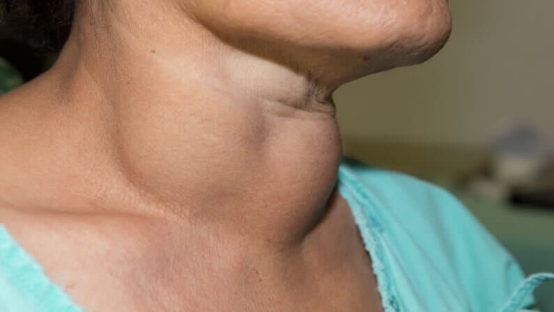
Pancreas Surgery in Turkey - Clinics, Doctors & Reviews
Discover the top clinics, best doctors, and various methods used in pancreatic surgery in Turkey through this article.

Liver Cirrhosis Treatment in Turkey - Best Clinics and Costs
This article is about the treatment of cirrhosis in Turkey. Check out our article for information about liver transplantation in Turkey for the treatment of cirrhosis, the best clinics and doctors, co...

Meningioma (Brain Tumor) Removal Surgery in Turkey
For those contemplating meningioma surgery in Turkey but lacking knowledge about the process, this article serves as a valuable resource for obtaining essential information.
Our doctor is highly skilled and an expert in their field.


